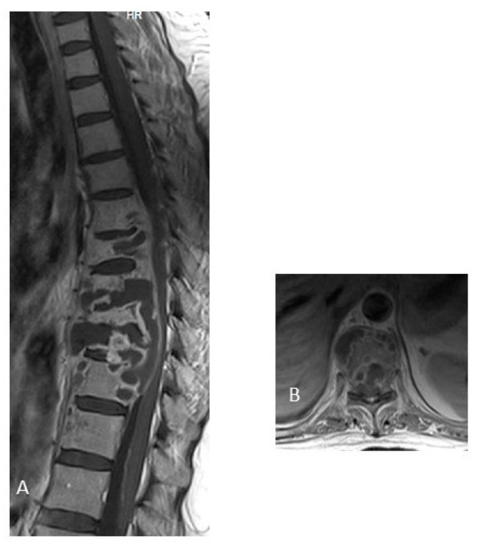
Figure 4.
MRI of thoracic spine post-contrast T1-weighted image. (A) Sagittal image, and (B) Axial image, with lytic lesions of the vertebral bodies and appendiceal involvement from T7 to T9, with prevertebral and epidural abscess. A 29-year-old woman, with pulmonary tuberculosis, who had low back pain for 14 months, spinal tissue culture positive for M. tuberculosis. The image shows multilevel tuberculous spondylodiscitis from T5 to T11 with disc-vertebral destruction with epidural abscess with compression of the spinal cord from T7 to T10. (The photo is from our image gallery).

