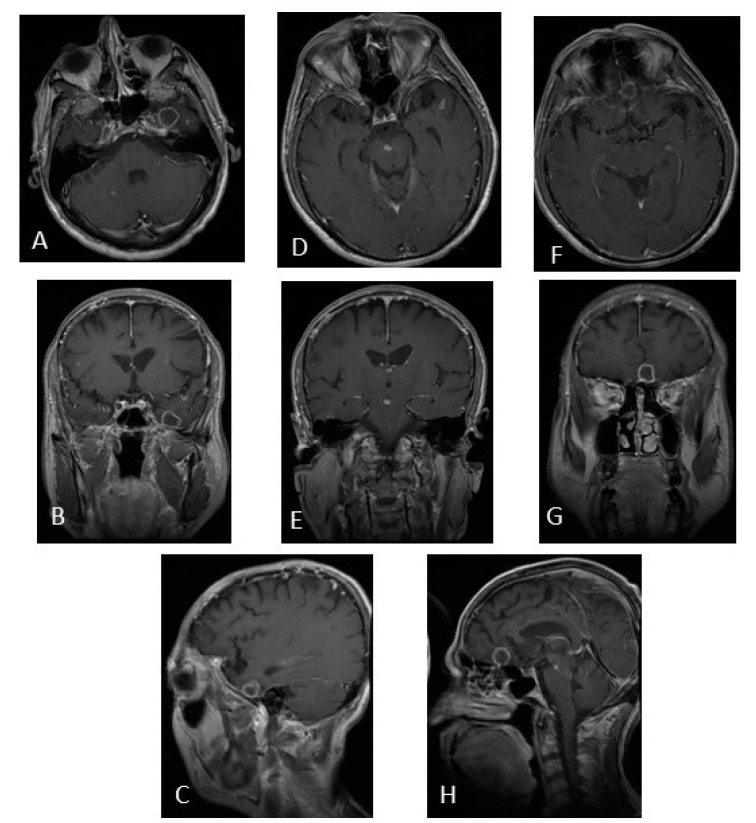
Figure 5.
MRI of brain post-contrast T1-weighted image. (A–C) Tuberculoma in the left temporal lobe. (D,E) Tuberculoma in the midbrain. (F–H) Tuberculoma in the frontal lobe. Sixty-four-year-old patient, with previous use of infliximab, with a fever of 6 months of evolution. Clinical improvement at 2 weeks, and imaging improvement at 9 months after starting antituberculous drugs. (The photo is from our image gallery).

