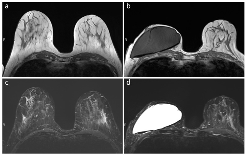
Figure 1.
T1-weighted (panels (a,b)) and T2-weighted (panels (c,d)) MRI images of a patient that underwent a skin sparing mastectomy of the right breast with pre-pectoral implantation of the MRI-conditional breast tissue expander. (Panels (a,c)) show preoperative MRI images, while (panels (b,d)) show MRI images acquired prior to planned exchange surgery to definitive implant, with no negative effects on image quality caused by the presence of the tissue expander and its non-metallic port.

