Figure 1.
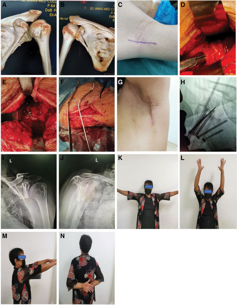
Female patient, 64 years old, was hit by a car while riding her electric bicycle. Preoperative CT 3D reconstruction (A, B) shows an Ideberg II scapular glenoid fracture with the scapular glenoid fracture line extending to the lateral edge of the scapula. The operation was treated by axillary approach with general anesthesia in the supine position, the incision was made from the apex of the axilla along the anterior border of the latissimus dorsi muscle towards the proximal end of the trunk to the level of the subscapular angle (C), and the axillary nerve and the posterior spinohumeral artery were revealed (D). The glenohumeral capsule is further exposed by blunt inward separation along the anterior border of the latissimus dorsi muscle gap, and the fracture is subsequently repositioned and fixed with screws (E). The Hohmann pulling hook was intraoperatively used to pull the muscle (F). Intraoperative X-ray fluoroscopy reveals a well-positioned scaphoid fracture with a hollow screw (H). The incision is closed with sutures, approximately 7 cm long (G), and the incision can be obscured when the affected limb drops. Positive and lateral x-rays of the scapula of the shoulder joint 3 months after surgery showed that the scapular pelvis fracture was well reset (I, J), and the patient’s shoulder function returned to normal (K–N).

