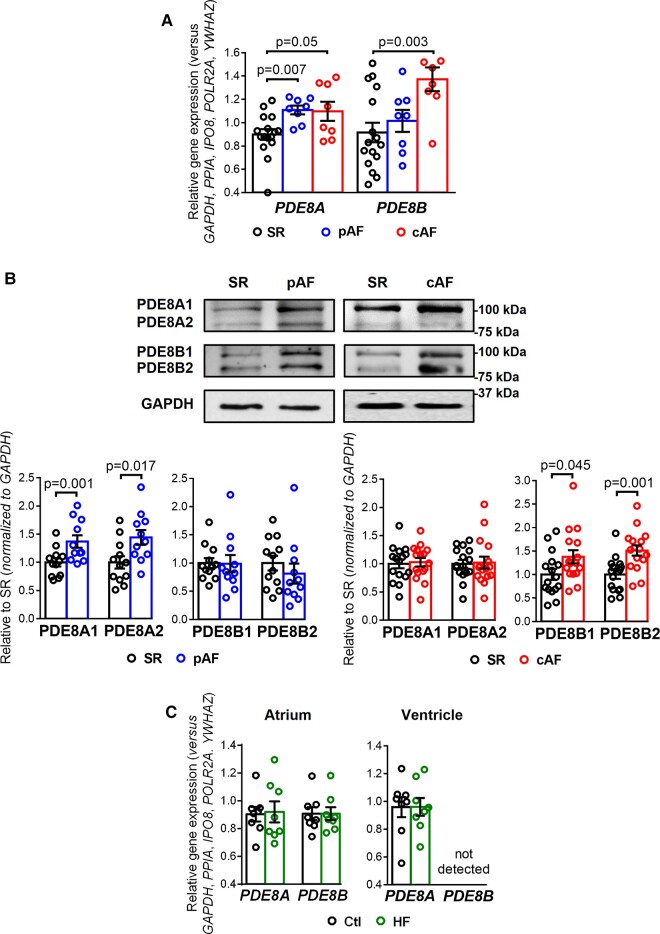
Figure 1.
PDE8A and PDE8B expression in human atrium. (A) Plot of the individual and mean ± SEM gene expression ratio of PDE8A and PDE8B normalized to a set of five housekeeping genes (POLR2A, YWHAZ, GAPDH, IPO8, PPIA) in atrial tissue homogenates from 16 sinus rhythm (SR), 8 paroxysmal atrial fibrillation (pAF) and 8 persistent (chronic) atrial fibrillation (cAF) patients. *P < 0.05 vs. sinus rhythm SR based on analysis of variance (ANOVA with a Kruskal–Wallis test). (B) Representative western blot (top) and protein expression quantification (bottom, mean ± SEM) of PDE8A and PDE8B in atrial tissue homogenates from 16 SR, 10 pAF and 16 cAF patients. GAPDH was used as loading control. *P < 0.05 vs. SR based on unpaired Student’s t-test analysis. (C) Plot of the individual and mean ± SEM gene expression ratio of PDE8A and PDE8B normalized to the set of five housekeeping genes in right atrial and left ventricular tissue homogenates from eight healthy control (Ctl) and eight end-stage heart failure (HF) patients. *P < 0.05 vs. Ctl based on ANOVA and Mann–Whitney test.

