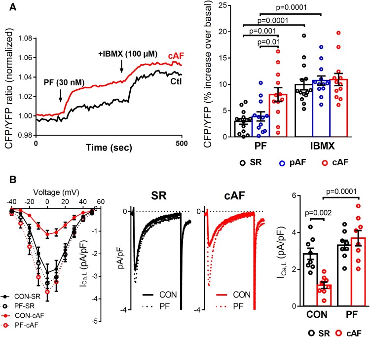
Figure 4.
Functional consequences of selective PDE8 inhibition in human atrial myocytes (HAMs). (A) Left panel, representative experiments showing the time course of the FRET signals indicating cAMP changes in HAMs from sinus rhythm (SR) and in persistent (chronic) atrial fibrillation (cAF) patients exposed to selective PDE8 inhibition with PF-04957325 (30 nM). Right panel, effects of the selective PDE8 inhibitor and the non-selective PDE blocker IBMX (100 mM) on the individual and mean values of cAMP levels expressed as percentage of increase of CFP/YFP over its control value measured in 14 HAMs from 6 SR, 12 HAMs from 6 paroxysmal AF (pAF), and 12 HAMs from 6 cAF patients. The changes in FRET ratio were expressed as a percentage change vs. basal. *P < 0.05 compared with SR, # P < 0.05 compared with PF. (B) Left panel, current-voltage (I-V) relationship for peak inward ICa,L density in HAMs from SR and cAF patients before (CON) and after exposure to PF. Middle panels, representative L-type Ca2+ current (ICa,L) recordings measured at 0 mV in HAMs from SR and cAF patients before (CON) and after exposure to PF. Right panel, average current densities before and after exposure to PF (n = 8 cells/5 patients SR and 8/5 cAF). *P < 0.05 vs. sinus rhythm SR, # P < 0.05 vs. CON. Statistical differences analyzed by mixed ANOVA followed by Wald χ2 test and Sidak multiple comparison test.

