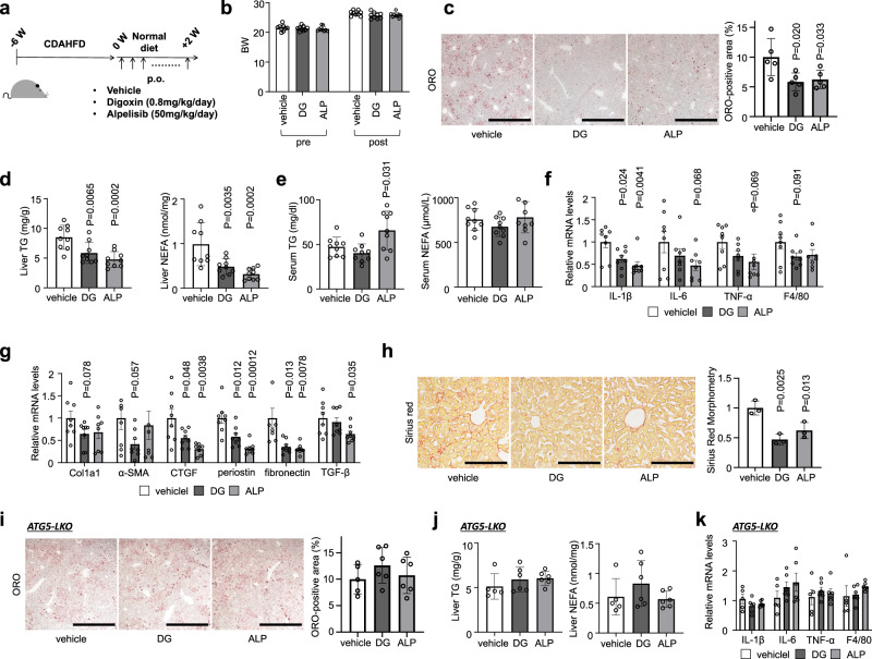
Fig. 7. Alpelisib and digoxin prevented the progression of NASH in mice fed CDAHFD.
a Study protocol of digoxin (DG) or alpelisib (ALP) administration in NASH model mice with CDAHFD. b Body weight of control and drugtreated mice. c ORO staining of liver sections. Scale bars, 1 mm. d Liver TG and NEFA content. e Serum TG and NEFA levels. f, g Real-time PCR assessing the expression of inflammation- (f) and fibrosis- (g) related genes in liver tissue. h Sirius red staining of liver sections. Scale bars, 200 µm. i ORO staining of liver sections of liver-specific Atg5 knockout mice fed CDAHFD. j Liver TG and NEFA content of liver-specific Atg5 knockout mice fed CDAHFD. k Real-time PCR assessing the expression of inflammation-related genes in Atg5 knockout liver tissue. Data are presented as mean ± SD of n = 8–9 (a–h) and n = 5–6 (i–k). P values calculated by one-way ANOVA with Dunnett’s multiple comparison test. Source data are provided as a Source data file.

