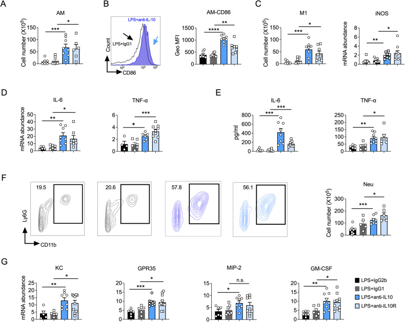
Fig. 2. Interleukin (IL)-10 signaling blockade leads to exacerbated lung inflammation after lipopolysaccharide (LPS) exposure.
A Flow cytometry analysis of macrophage cell numbers in mice lung (n = 6–8). B Geo MFI of CD86 on macrophages. (n = 6–8). C Flow cytometry analysis of M1 macrophage numbers in mice lung (left); RT-PCR to test the expression level of iNOS mRNA (right) (n = 6–10). D The mRNAs of IL-6 and TNF-α were detected in lung homogenates (n = 4–10). E Cytokine levels in bronchoalveolar lavage fluid samples of different groups (n = 5–9). F Neutrophils (CD11b+, Ly6G+) in the lungs as a proportion of live events. The total number of cells was then multiplied by the ratio to give the total neutrophil count. (n = 5–9). G Chemokines associated with neutrophils (n = 5–11). A–G Experiments were conducted 4 days after LPS exposure. A–G Student’s t-test was used for the statistical analyses.

