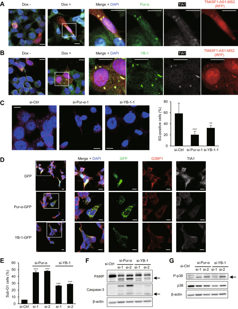
Fig. 7. Pur-α and YB-1 contribute to SG formation and apoptosis inhibition in GC cells.
A, B Immunofluorescence images showing Pur-α (A) or YB-1 (B), TIA1 and inducible MS2-tagged TM4SF1-AS1 in SNU638 cells incubated for 8 days with or without Dox. Scale bars = 10 μm. C Immunofluorescent staining of G3PB2 in SNU638-TM4SF1-AS1 cells transfected with the indicated siRNAs. Representative results are shown on the left. Summarized results are shown on the right. (n = 5). Scale bars = 10 μm. D Localization of GFP-tagged Pur-α or YB-1, G3BP1 and TIA1 immunofluorescence in SNU638 cells. Cells were transfected with vectors encoding GFP (upper), GFP-tagged Pur-α (middle) or GFP-tagged YB1 (bottom). Magnified views of the respective markers are shown on the right. Scale bars = 10 μm. E Cell cycle analysis of HSC-45 cells expressing the indicated siRNAs. (n = 3). F, G Western blot analysis of PARP and caspase-3 (F) and total and phosphorylated p38 (G) in HSC-45 cells transfected with the indicated siRNAs. Cleaved PARP, cleaved caspase-3 and phosphorylated p38 are indicated by arrows. **P < 0.01, ***P < 0.001.

