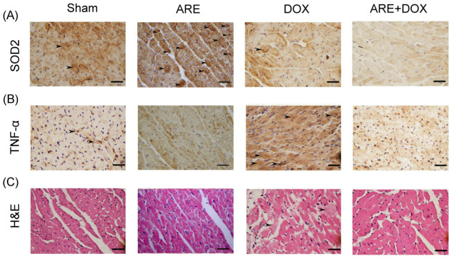
Figure 6.
Effect of ARE treatment on myocardial tissue of BALB/c breast cancer mice with DOX chemotherapy: (A) Myocardial IHC expression of SOD2 (marked with arrows) among BALB/c breast cancer mice with Sham, ARE (150 mg/kg), DOX (2.4 mg/kg), and ARE (150 mg/kg) + DOX (2.4 mg/kg) treatments. SOD2 expression was obviously reduced after DOX chemotherapy and ARE+DOX treatments. (B) Myocardial IHC expression of TNF-α (marked with arrows) in the BALB/c breast cancer mice was obviously enhanced after DOX chemotherapy but obviously reduced after ARE+DOX treatments. (C) H&E expression showed myocardial tissue damage (marked with arrows) in the BALB/c breast cancer mice after DOX chemotherapy; however, this was not shown in the BALB/c breast cancer mice after ARE+DOX treatments. Scale bar = 30 μm.

