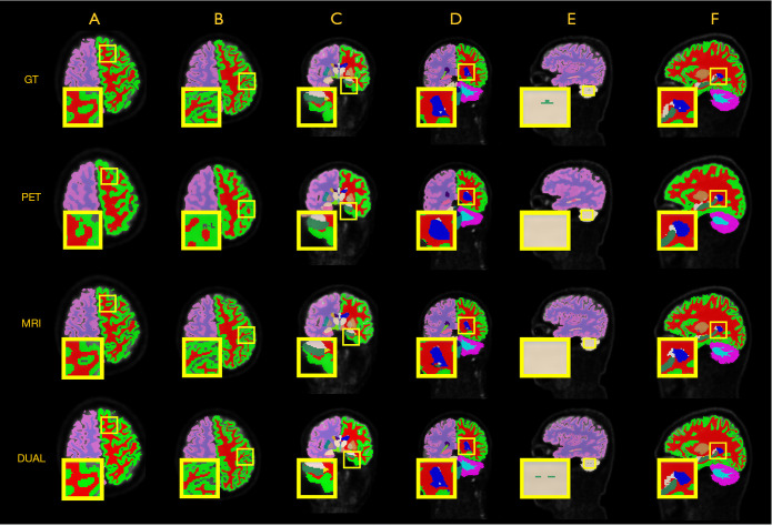
Figure 4.
Segmentation results of all classes shown in axial, coronal, and sagittal views. The first row shows the ground truth, and the second row presents results from the PET-only-based segmentation, while the third row presents the results from the MRI-only-based segmentation. The last row shows the results of our method, and the yellow boxes denote the regions of interest. (A) Frontal lobe; (B) parietal lobe; (C) temporal occipital lobe; (D) lateral ventricle; (E) cerebellum; (F) corpus callosum. DUAL, dual-modality method; GT ground truth; MRI, magnetic resonance imaging; PET, positron emission tomography.

