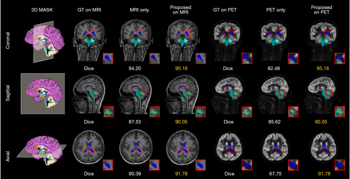
Figure 8.
Segmentation results of the left lateral ventricle, left cerebellum white matter, right lateral ventricle, right cerebellum white matter and brain stem in the axial, coronal, and sagittal views. The Dice values are the mean values of the slices for the given labels, and the best results are marked in yellow. GT, ground truth; MRI, magnetic resonance imaging; PET, positron emission tomography.

