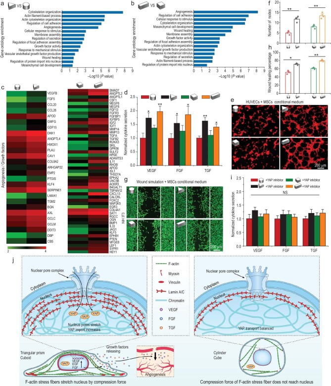
Figure 5.
3D micropattern mechanical force regulates MSCs paracrine function by YAP nuclear localization. (a and b) Gene ontology analysis of significantly regulated genome in 3D micropatterned cells. (c) Gene chip heatmaps for the differential genes related to angiogenic factor and growth factor in 3D micropatterned cells. (d) ELISA measurement of the paracrine products of pro-angiogenic and pro-regenerative factors secreted by MSCs cultured in 3D micropatterns. (e) Tube formation images of HUVECS incubated with paracrine products from different 3D micropatterned MSCs for 6 h. (f) Quantitative analysis of tube formation assay. (g) Representative images of NIH 3T3 migration to the defined ‘artificial wound’ after treatment with conditional medium from different 3D micropatterned MSCs for 24 h. (h) Quantification of wound healing percent on NIH 3T3 cells by wound simulation scratch assay. (i) Effects of YAP inhibitor on the secretion of pro-angiogenic and pro-regenerative factors of 3D micropatterned MSCs detected by ELISA measurement analysis. The data are represented as the mean ± SD, * p < 0.05, ** p < 0.01. NS, no significant difference. (j) Schematic representation of mechanotransduction pathways of MSCs triggered by 3D micropatterns and its effect on paracrine function of MSCs.

