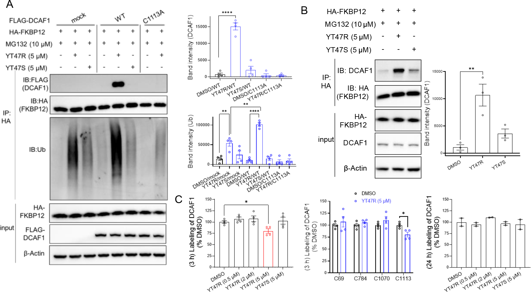
Figure 6. Evidence for ternary complex formation and ubiquitination of HA-FKBP12 by DCAF1-directed electrophilic PROTACs.

(A) Left, western blots showing effects of YT47R and YT47S on the immunoprecipitation (IP) of FLAG-DCAF1-WT or FLAG-DCAF1-C1113A with HA-FKBP12 in HEK293T cells co-expressing these proteins. Cells were treated with YT47R or YT47S (5 μM) for 2 h in the presence of MG132 (10 μM) prior to lysis and IP with an anti-HA antibody and western blotting for the indicated proteins (DCAF1, FKBP12, and ubiquitinated protein). Right, bar graphs quantifying DCAF1 (top) and ubiquitination (Ub) (bottom) in the indicated IP groups. (B) Left, western blots showing effects of YT47R and YT47S on the IP of endogenous DCAF1 with HA-FKBP12 in HEK293T cells expressing HA-FKBP12. Cells were treated with YT47R or YT47S (5 μM) for 2 h in the presence of MG132 (10 μM) prior to lysis and IP with an anti-HA antibody and western blotting for the indicated proteins (DCAF1, FKBP12). Right, bar graph quantifying DCAF1 in the indicated IP groups. For (A, B), data are mean values ± SEM for three independent experiments. Statistical significance was calculated with unpaired two-tailed Student’s t-tests comparing YT41R/YT47R- and DMSO-treated cells. **P < 0.01, ****P < 0.0001. (C) MS-ABPP quantification of IA-DTB labeling of the indicated cysteines in DCAF1 from FLAG-DCAF1-WT-expressing HEK293T cells treated with the indicated concentrations of YT47R for 3 h (left, middle) or 24 h (right) relative to cells treated with DMSO. Data represent average values ± SEM from four independent experiments. Statistical significance was calculated with unpaired two-tailed Student’s t-tests comparing compound to DMSO treated cells. *P < 0.1.
