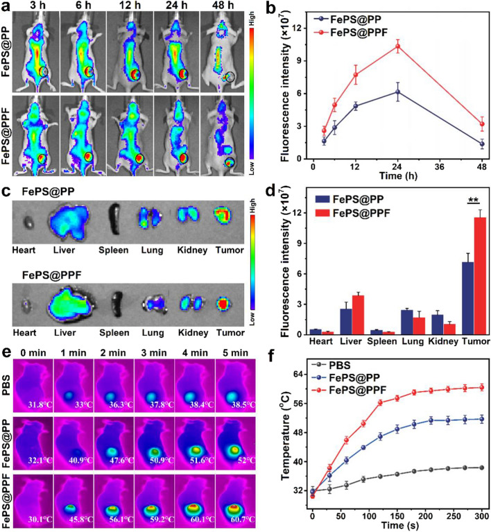
Fig. 4.
In vivo biodistributions of FePS@PPF. a In vivo fluorescence images of mice with HOS tumors (marked by ellipses) after injection of Cy5.5-labeled FePS NSs without (FePS@PP) or with folic acid modification (FePS@PPF). b Fluorescence intensity of tumor sites obtained by Living Image software. c Ex vivo fluorescence images. d Average fluorescence intensity of main organs and tumor, **p < 0.01. e Infrared thermographic images and f Temperature changes of tumor site under laser irradiation (1064 nm, 1.0 W/cm2) at 24 h after respectively intravenous infusion of PBS, FePS@PP and FePS@PPF

