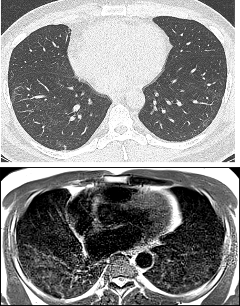
Fig. 5.
46 year old man with history of Covid 19 pneumonia 5 months prior to MRI. Right greater than left lower lobe groundglass opacity is apparent on CT (A). Low-field MRI obtained 2 days after CT demonstrates similar extent and distribution of groundglass opacity (B). Signal intensity on MRI appears to suggest greater severity of opacity than CT attenuation.

