Figure 5.

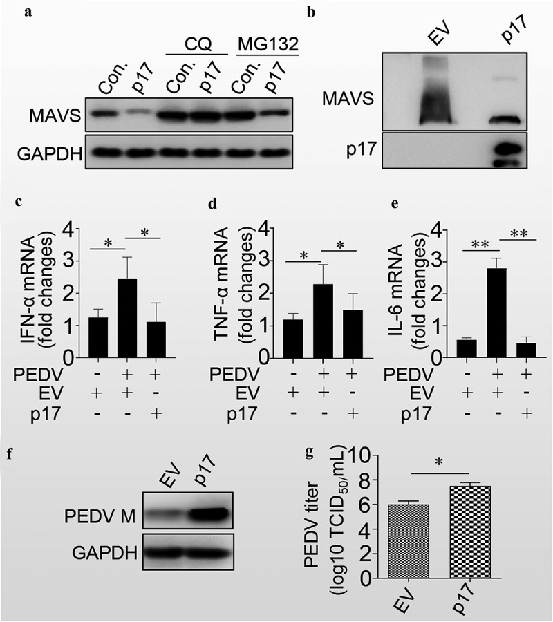
P17 inhibits the innate immune response by autophagic degradation of MAVS.
(a) HEK-293T cells were transfected with empty vector or vectors expressing MAVS, MDA5, and RIGI in the presence or absence of CQ or MG132. The cells were harvested for WB analysis by using anti-MAVS, MDA5, and RIGI antibodies. (b) HEK-293T cells were transfected with empty vector or vectors expressing Flag-p17. The cells were harvested for WB analysis by using anti-MAVS or anti-Flag antibodies. (c, d and e) VERO cells were transfected with empty vector or vector expressing p17 for 24 hours and were then infected with PEDV for 24 hours. The cells were then harvested for qPCR analysis. (f) Vero cells were transfected with empty vector or vector expressing p17 and were then harvested for WB analysis by using anti-PEDV M and GAPDH monoclonal antibodies. (g) Vero cells transfected with EV or vector expressing p17 were infected with PEDV, and the TCID50 was then tested. At least three independent experiments were performed. The error bars show the standard error of the mean (SEM). At least three independent experiments were performed. The error bars show the standard error of the mean (SEM). Significance was analysed with two-tailed Student’s test. *p<0.05, **p<0.01, ***p<0.001.
