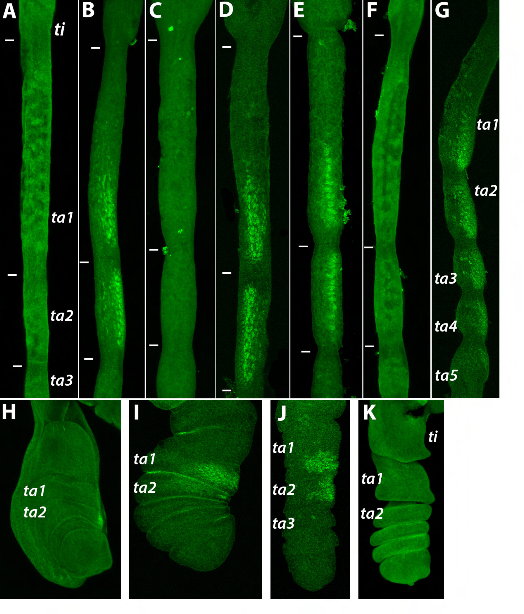
Figure 3. Dsx expression in Lordiphosa T1 legs.

Proximal is up, distal is down. Tibia (ti) and tarsal segments 1–5 (ta1–ta5) are indicated in some panels; in other panels, the boundaries between these segments are shown with white tick marks. Panels A–G show pupal legs at stages equivalent to 24–30 hours After Puparium Formation (APF) in D. melanogaster. A. L. acongruens male; Dsx protein is absent. B. L. clarofinis male; Dsx is expressed on the anterior-ventral leg surface in the distal part of ta1 and all of ta2. C. L. clarofinis female; no Dsx expression is seen at this stage. D. L. sp.1 male; Dsx expression is similar to L. clarofinis. E. L. sp.2 male; Dsx expression is similar to L. clarofinis. F. L. sp.3 male; no Dsx expression is observed. G. L. collinella male; Dsx staining is present in all tarsal segments. H. The T1 leg imaginal disc of the white prepupa (0 hrs APF) of L. acongruens male; no Dsx expression is observed. I. Prepupal leg of L. sp.2 male at the stage equivalent to ~2–3 hrs APF in D. melanogaster; Dsx is expressed on the anterior-ventral surface of ta1 and ta2. J. Prepupal leg of L. sp.2 male at the stage equivalent to ~6–7 hrs APF in D. melanogaster. K. Prepupal leg of L. sp.3 male at the stage equivalent to ~5–6 hrs APF in D. melanogaster; no Dsx expression is seen.
