Figure 4.

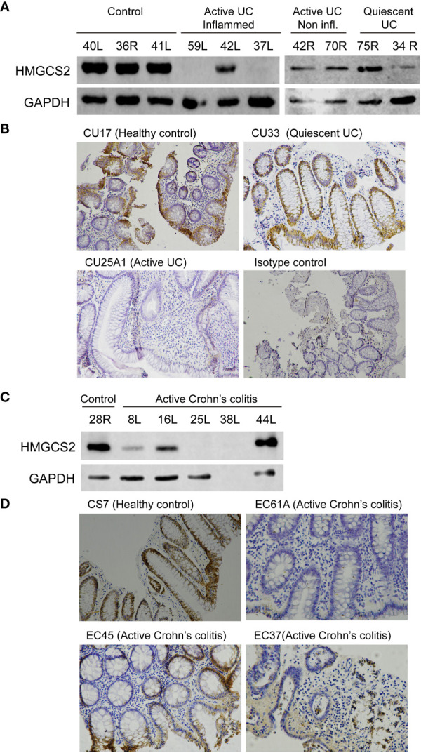
HMGCS2 protein expression is decreased in colonic samples from IBD patients. (A) Representative immunoblot of HMGCS2 expression in colonic samples from UC patients with active or quiescent disease or healthy controls compared to GAPDH expression. (B) Representative HMGCS2 immunohistochemistry using paraffined-tissue slides. Samples CU17 (Healthy control) and CU33 (Quiescent UC) had positive staining for HMGCS2, whereas CU25A1 (Active UC) displayed no staining for HMGCS2. Sample CU33 (Isotype control) was used as a negative control. (C) Representative immunoblot of HMGCS2 expression in colonic samples from healthy controls and patients with active colonic Crohn’s disease. (D) Representative HMGCS2 immunohistochemistry of active Crohn’s colitis biopsies (EC61A and EC37) which showed no staining, whereas healthy control biopsy (CS7) and active Crohn’s colitis biopsy (EC45) were positive for HMGCS2 staining.
