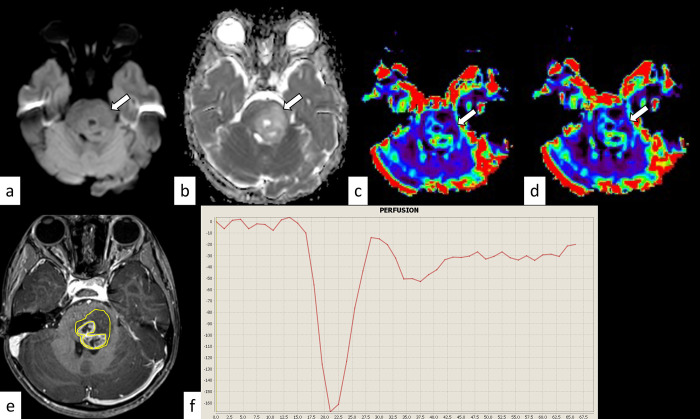
Fig 3. H3.1 variant diffuse midline glioma, H3K27-altered in a 4-year-old girl.
The mass shows isointensity on diffusion-weighted imaging (a, arrow) and an increased apparent diffusion coefficient (nADCmean = 1.72) (b, arrow). The normalized relative cerebral blood volume and flow are 1.76 and 1.56, respectively (c and d, arrows). The corresponding post-contrast T1-weighted image and region-of-interest in this slice are shown (e). The percentage signal recovery of the mass is 0.78 (f). nADCmean = normalized mean apparent diffusion coefficient.

