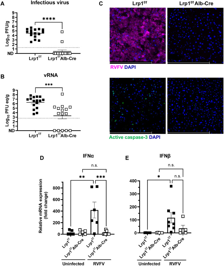
Fig. 3. Lrp1 KO in hepatocytes leads to a significant reduction in viral burden in the liver at 3 dpi.
(A) Lrp1f/fAlb-Cre mice (n = 16) have significantly less (A) infectious virus and (B) viral RNA (vRNA) in the liver at 3 dpi than Lrp1f/f controls (n = 15). (C) RVFV nucleoprotein (N) (magenta) and active caspase-3 (green) staining in the liver at 3 dpi in Lrp1f/f and Lrp1f/fAlb-Cre livers (40×). (D) Interferon α (IFNα) and (E) IFNβ relative mRNA expression in the liver of uninfected and RVFV-infected Lrp1f/f and Lrp1f/fAlb-Cre mice at 3 dpi. Bars represent SEM. Statistics were determined by an unpaired t test or a one-way analysis of variance (ANOVA). *P < 0.05; **P < 0.01; ***P < 0.001; ****P < 0.0001. n.s., not significant. Scale bars, 250 μm. Dotted lines indicate the limit of detection.

