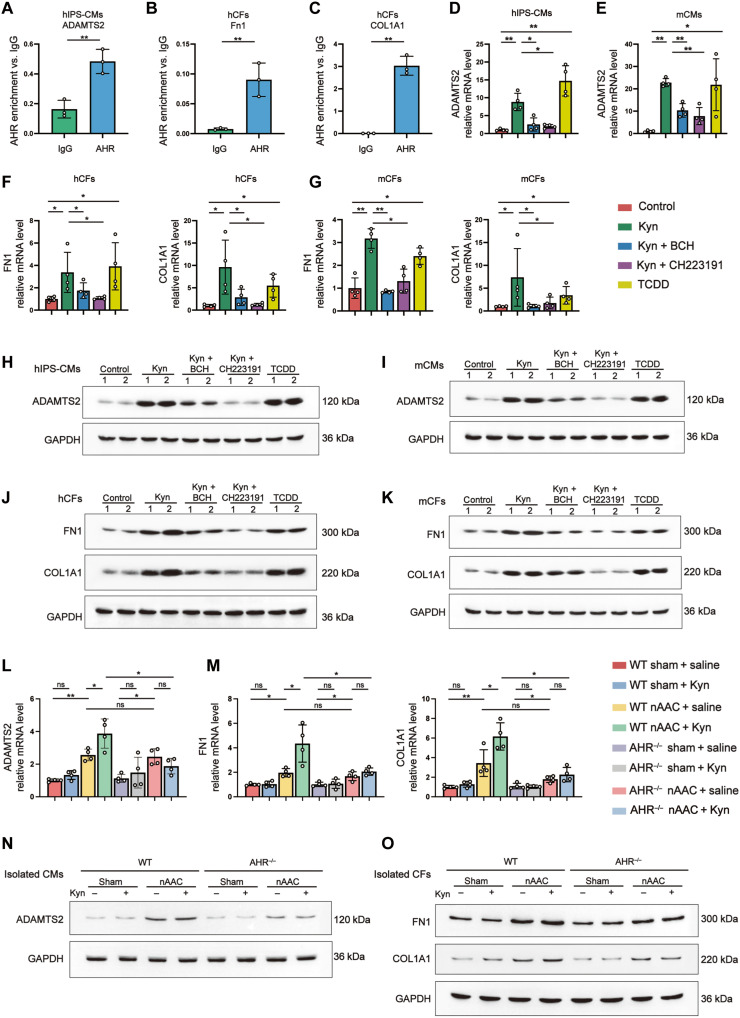
Fig. 4. Kyn-AHR activation initiated the expression of hypertrophy and fibrosis genes.
(A to C) AHR enrichment in comparing to immunoglobulin G in chromatin immunoprecipitation of ADAMTS2 in hIPS-CMs (A), FN1 (B), and COL1A1 (C) in hCFs. n = 3 per group. (D and E) qPCR quantified mRNA level of ADAMTS2 in hIPS-CMs (D) and mCMs (E), treated for 48 hours with 500 μM Kyn, 500 μM Kyn and 500 μM BCH, 500 μM Kyn and 500 μM AHR inhibitor CH223191, 500 μM TCDD as positive control, or the same amount of DMSO as negative control. (F and G) mRNA level of FN1 and COL1A1 in hCFs (F) and mCFs (G) treated with indicated compounds. (H and I) Representative Western blotting of the protein expression ADAMTS2 in hIPS-CMs (H) and mCMs (I) treated with indicated compounds. Each lane represented individual experiment in vitro. (J and K) Representative Western blotting of FN1 and COL1A1 in hCFs (J) and mCFs (K) treated with indicated compounds. Each lane represented individual experiment in vitro. (L and M) qPCR quantified mRNA level of ADAMTS2 in isolated mCMs (L) and FN1 and COL1A1 in isolated mCFs (M) from WT and AHR−/− mice 4 weeks after sham or nAAC surgery, with Kyn peritoneal injection (50 mg/kg) or saline as control. (N and O) Representative Western blotting of the protein expression of ADAMTS2 in isolated mCMs (N) and FN1 and COL1A1 in isolated mCFs (O) from indicated mice. Each lane represented individual animal. Two-tailed Mann-Whitney U test was used in (A) to (C). Kruskal-Wallis test and post hoc Dunn test were used in (D) to (G), (L), and (M). *P < 0.05 and **P < 0.01.

