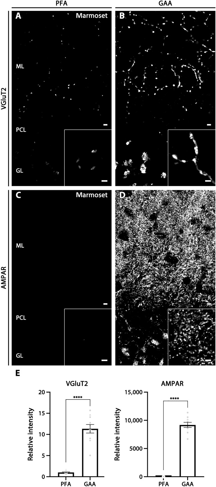
Fig. 2. Improved immunostaining by GAA fixation in the cerebellar cortex of marmoset brains.
(A and B) VGluT2 labeling in PFA- (A) or GAA-fixed (B) cerebellum. (C and D) AMPAR labeling in PFA- (C) or GAA-fixed (D) cerebellum. Insets show enlarged images of the cerebellar molecular layer. (E) Histogram showing the mean relative ratio in GAA-fixed sections normalized to PFA-fixed sections in the cerebellar molecular layer (10 images from two marmosets). For detailed statistics, see Table S2 and S3. For abbreviations, see Fig. 1. Scale bars, 5 μm (A to D) (inset, 2 μm).

