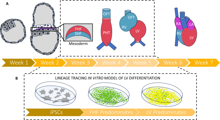
Figure 1. A new strategy for tracing the fate of iPSCs as they develop into muscle cells of the heart.
(A) Timeline (in weeks post-fertilization) of early human embryonic heart development from the blastocyst through to the emergence of the mesoderm and first (red) and second (blue) heart fields. These each give rise to different parts of the four-chambered heart: the first heart field will go on to form the primitive heart tube and left ventricle, while the second heart field cell will go on to form the right ventricle and the outflow tract. The period of development between two and five weeks post fertilization (depicted in inset) remains understudied in humans due to the 14day rule governing how long human embryos can be grown in the laboratory, as well as the low availability of fetal and embryonic tissues that are less than six weeks post-fertilization. (B) Galdos et al. developed a robust lineage tracing strategy to evaluate iPSCs that had been differentiated into cardiac cells by modulating the Wnt pathway. Under this approach, cells transitioning into first heart field cells fluoresce green, and those that terminally differentiate into ventricle muscle fluoresce red. Most cells displayed both fluorescent proteins (resulting in a yellow color), suggesting that Wnt-based differentiation predominately generates muscle cells of the left ventricle. Abbreviations: FHF – first heart field; SHF – second heart field; OFT – outflow tract; PHT – primitive heart tube; RV – right ventricle; LV – left ventricle; RA – right atrium; LA – left atrium; iPSC – induced pluripotent stem cell.

