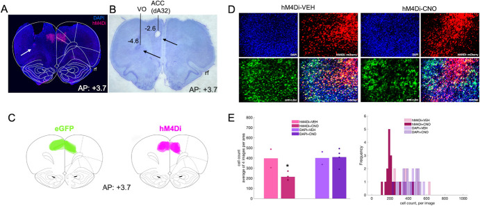
Figure 3.
Inhibitory DREADDs in ACC, electrode placement in ACC and OFC, and validation of inhibition in ACC via c-fos immunohistochemistry. (A) Representative placement of inhibitory hM4Di DREADDs at anterior–posterior (AP) level + 3.7 relative to Bregma. Note, tissue (secondary motor cortex, M2) was removed at time of brain extraction. White arrow pointing at tip of electrode track in OFC. (B) Nissl-stained section showing electrolytic lesions and placement of electrode arrays targeting both ACC (dorsal area 32, dA32) and OFC (ventral orbital, VO) unilaterally in a representative animal. Black arrows pointing at tip of electrode tracks. Rf = rhinal fissure. (C) Reconstructions of placement of inhibitory hM4Di DREADDs (right) and eGFP null virus (left) at anterior–posterior (AP) level + 3.7 relative to Bregma. (D) Representative DAPI, hM4Di-mCherry, c-fos immunoreactivity and their overlap after injections of VEH (left) or CNO (right). (E) Mean cell count of 4 images per condition, hM4Di + VEH, hM4Di + CNO, DAPI+VEH, and DAPI+CNO. One-way ANOVA resulted in a significant decrease in c-fos–positive cells in the hM4Di-expressing areas, but not in the DAPI areas (left). Histogram of cell count frequency for each image by condition (right). *P < 0.05.

