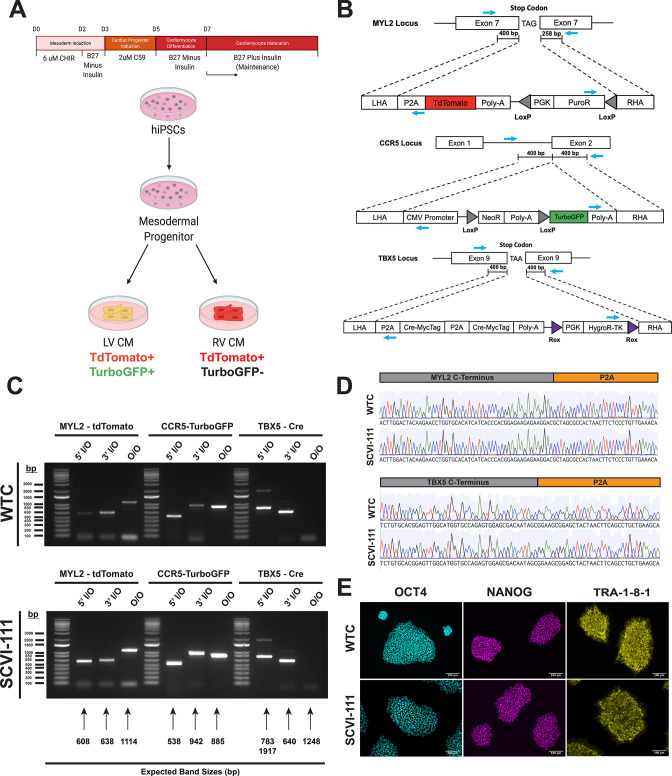
Figure 1. Integration of T-box transcription factor (TBX5)/myosin light chain-2 (MYL2) lineage tracing reporter system into human-induced pluripotent stem cells.
(A) Schematic of lineage tracing strategy for identifying left ventricular cardiomyocytes in vitro. (B) CRISPR/Cas9 gene targeting strategy of genetic constructs for TBX5 lineage tracing and MYL2 direct reporter. MYL2, CCR5, and TBX5 constructs contain Puromycin (PuroR), Neomycin (NeoR), and Hygromycin (HygroR) resistance cassettes for the selection of human-induced pluripotent stem cell (hiPSC) after targeting of genetic constructs. Blue arrows indicate the location of PCR primer binding sites for confirmation of construct integration. LHA = Left Homology Arm, RHA = Right Homology Arm. (C) Inside-Outside (I/O) and Outside-Outside (O/O) PCR DNA agarose gels for confirmation of integration of genetic constructs into MYL2, CCR5, and TBX5 genetic loci. Inside, represents a primer inside the construct region while Outside represents a primer that binds outside the homology arm regions of genetic constructs. Expected band sizes are noted with arrows for each lane. (D) Sanger sequencing traces for C-terminal regions of MYL2 and TBX5 genes indicating in-frame integration of P2A site. (E) Bright field and immunofluorescence images of pluripotency marker expression in hiPSC lines after integration of all three genetic constructs.


