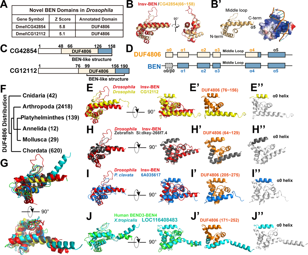
Figure 3. The DUF4806 motif represents a subgroup of BEN domain with an upstream α0 helix.
(A) Structure comparison shows Drosophila CG42854 and CG12112 to be similar to BEN domains, with BEN-like regions overlapping with a presumed DUF4806 motif.
(B-B') Superposition of the Insv-BEN structure (PDB: 4IX7, red) and the predicted model of CG42854. (B') shows the electrostatic surface potential of the CG42854 BEN-like domain with Insv-BEN targeting DNA.
(C) The domain organization of Drosophila CG42854 and CG12112 proteins, with BEN-like structures overlapping with uncharacterized DUF4806 motifs.
(d) A schematic view of DUF4806 and BEN domains. DUF4806 contains five α-helices (α0-α4) preceding a downstream unrecognized helix α5, and the α1-α4 helices in BEN domains correspond well with DUF4806. In contrast, there could be either α-helices (BEND3-BEN3, PDB:7V9H) or β-sheets (Insv-BEN, PDB: 4IX7) at the upstream of BEN domains.
(E-E”) Superposition of Insv-BEN (PDB: 4IX7, red), and the predicted structure of CG12112 BEN-like region, showing that DUF4806 closely resemble Insv-BEN domains. (E’) shows the cartoon view of predicted structures of CG12112 regions containing α0-α5 helices, with residues corresponding to DUF4806 colored in orange. Note that DUF4806 ends in the middle of helix α4. (E”) shows the α0-α5 helices of CG12112, with core BEN domain structure (α1-α5 helices) colored in white and helix α0 highlighted with yellow.
(F) Phylum distribution of annotated DUF4806-containing proteins in the InterPro database.
(G-J) Superposition of Insv-BEN (PDB: 4IX7, red), BEND3-BEN4 (PDB: 7W27, green), and predicted DUF4806 models, showing that DUF4806 modules closely resemble solved BEN domains.
(H'-J') Cartoon view of predicted structures of regions containing α0-α5 helices, with residues corresponding to annotated DUF4806 colored in orange. Note that DUF4806 ends in the middle of helix α4.
(H''-J'') Cartoon view of predicted structures of regions containing α0-α5 helices, with core BEN domain structure (α1-α5/6 helix) colored in white and helix α0 highlighted with corresponding colors.

