Fig. 1.
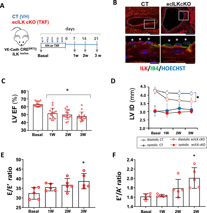
ILK deletion in coronary endothelium leads to myocardial dysfunction. A Schematic figure showing the experimental design. VE-Cadh-CreERT2/ILKlox/lox mice were treated (i.p.) during 5 days with Tamoxifen;TXF resulting in endothelial ILK deletion (ecILK cKO in red) or vehicle;VH (CT in blue). Cardiovascular function was tested before treatment (basal) and once a week up to three weeks. Hearts were harvested at 1, 2 and 3 weeks. B Representative confocal images of a coronary artery in CT (left panel) and ecILK cKO mice at three weeks stained with anti-ILK (red), endothelial cells were stained with IB4 (green) and nuclei were counterstained in blue with Hoechst. Asterisks in magnification indicate individual endothelial cells showing no expression of ILK (red) in the tamoxifen treated group. Scale bar (upper panel): 25 mm and 10 μm lower panel. C Bar graph showing the ejection fraction of the left ventricle (LV EF) in ecILK cKO mice (n = 15, *p < 0.001 vs Basal). D Changes in the internal diameter of left ventricle in diastole (LVIDd) and systole (LVIDs) in CT and ecILK cKO mice. (n = 15, *p < 0.005 vs CT diastole at three weeks). E. E/Eʹ ratio (LV end–diastolic filling pressure) and F Eʹ/Aʹ ratio from ecILKcKO mice (n = 5, *p < 0.001 vs Basal). All p values were calculated using ANOVA

