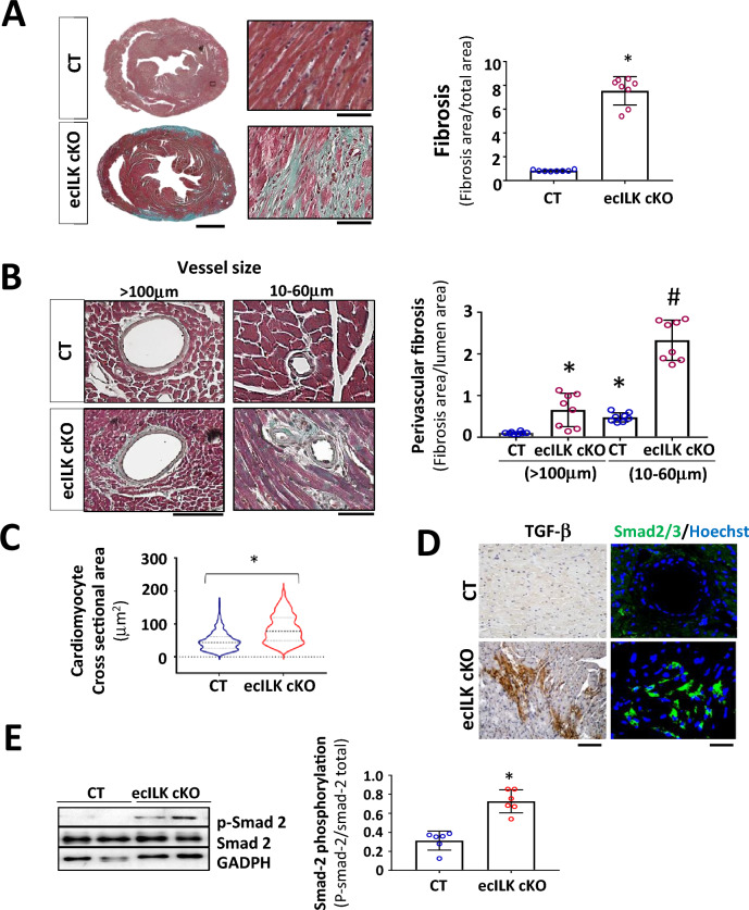
Fig. 2.
Endothelial-specific ILK deletion leads to adverse remodelling. A Representative Masson Trichrome staining of CT and ecILK cKO mice heart sections (Scale bar = 1 mm) showing cardiac lesions three weeks after deletion. Right: Magnification of ecILK cKO and CT hearts. Scale bar: 50 μm. Right panel: quantitative analysis of cardiac fibrosis. (n = 8,*p < 0, 005 vs CT). B Representative photo-micrographs from ecILK cKO and CT hearts stained with Masson Trichrome showing perivascular fibrosis in myocardial arteries > 100 μm diameter and small arterioles (60–10 μm). Scale bar: 100 μm (left panel); 50 μm (right panel). Right, quantitative analysis of perivascular fibrosis in vessels diameter > 100 μm and small arterioles (60–10 μm). (n = 8,*p < 0.001 vs CT > 100 μm; #p < 0, 005 vs CT small vessels). C Quantitative data of cardiomyocyte (CM) cell surface area; n = 6–10 hearts per group with 300–600 CMs analysed per heart. CM area is expressed in μm2. *p < 0, 05 CT vs ecILK cKO. D Representative heart sections of CT and ecILK cKO mice staining of TGF-β1 and SMAD2/3 (n = 8). Scale bar: 50 μm left and 25 μm right. E Immunoblot detection of phosphorylated Smad-2 in total heart lysates from CT and ecILK cKO mice. GAPDH was used as a loading control. Densitometric analysis (right panel) shows a significant increase in the expression of p-Smad2 in ecILK cKO mice (n = 6, *p < 0.05 vs CT). p values were calculated using Student’s t test and one-way ANOVA in B

