Fig. 3.
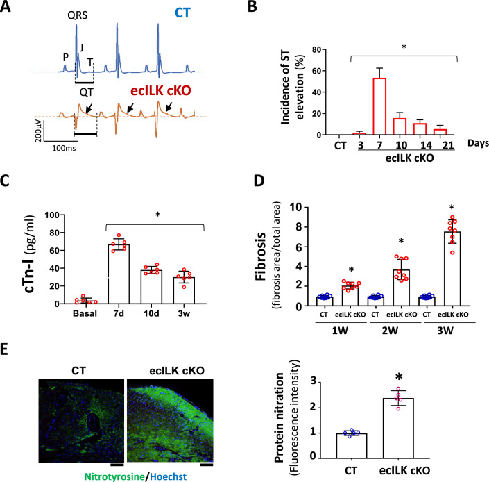
ILK deletion in coronary endothelium promotes myocardial ischaemia. A Representative ECG recording (lead II) of CT (upper panel) and ecILK cKO mice (lower panel) 1 week after treatment, showing ST elevation (arrow) in ecILK cKO mice. B Incidence of ST elevation in CT (21d) and ecILK cKO mice at different time points post-deletion in six different experimental groups (5–8 mice/group;*p < 0,001 vs CT). C Plasmatic cardiac troponin quantitation showing a significant increase 7 days after treatment when compared to basal conditions and remain elevated up to three weeks after deletion (n = 6 mice per condition; *p < 0.001 vs Basal). D Myocardial Fibrosis from CT and ecILK cKO mice 1, 2 and 3 weeks after deletion/treatment (n = 8 mice per condition) *p < 0.05 vs CT at the same time point. E Representative heart sections from CT and ecILK cKO mice 3 weeks after deletion stained with nitrotyrosine (green), nuclei were counterstained with Hoetchst (scale bar: 50 μm). Right panel, quantitative analysis showed a significant increase in protein nitration expressed as relative fluorescence intensity in ecILK cKO mice (n = 5, *p < 0.05). p values were calculated using one-way ANOVA and Student’s t test in (E)

