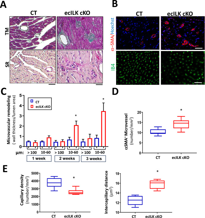
Fig. 4.
ILK endothelial deletion leads to altered microvascular structure and density. A Representative photomicrographs of heart sections stained with Masson Trichrome (upper panel) and Sirius Red (lower panel) showing the remodelled microvasculature in ecILK cKO (right panel) as compared to CT mice (left panel). Scale bar = 25 μm. B Representative photomicrographs of heart sections obtained from CT and ecILK cKO mice showing (upper panel) arterioles stained for smooth muscle actin (α-SMA, red) and counterstained in blue with Hoechst and (lower panel) capillaries stained for IB4 (green) Scale bar: 100 mm (n = 8). C Arteriolar wall thickness-to-lumen area ratio was significantly increased in ecILK cKO mice 2- and 3-weeks post-deletion only in small vessels (10–60 mm) and remained unchanged in large vessels (> 100 mm) (n = 8, *p < 0.05 vs CT). D Total number of α-SMA-positive microvessels (10-60 μm) was significantly increased in ecILK cKO mice three weeks after deletion (n = 6, *p < 0.05 vs CT). E In ecILK cKO mice, a significant decrease in capillary density (left panel) together with a significant increase in intercapillary distance (right panel) was observed 3 weeks after treatment when compared to CT mice (n = 6, *p < 0.05 vs CT). p values were calculated using Student’s t test and one-way ANOVA in (C)

