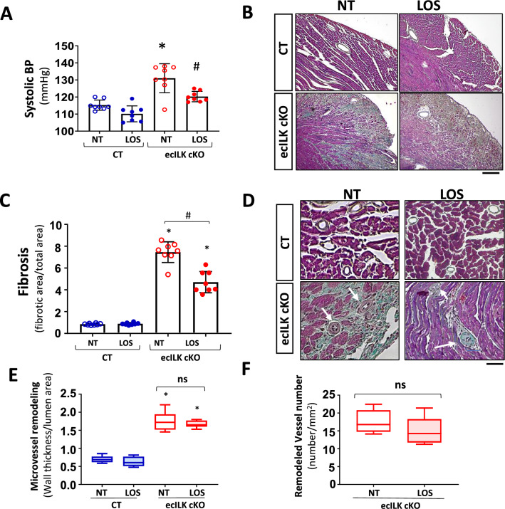
Fig. 5.
Effect of the antihypertensive drug Losartan, on the myocardial and microvascular remodelling of ecILK cKO mice. CT and ecILK cKO mice were treated with Losartan (LOS, n = 8) or left untreated (NT, n = 8). A Three weeks after treatment, systolic arterial blood pressure was significantly increased in untreated ecILKcKO as compared to CT mice; this increase was prevented by LOS (*p < 0.05 vs CT; # p < 0.05 vs NT ecILK cKO). B Representative photomicrographs of heart sections obtained from CT and ecILK cKO mice treated as in A, stained with Masson Trichrome showing myocardial fibrosis at three weeks (n = 8) Scale bar: 100 μm. C Quantitative analysis of myocardial fibrosis at 3 weeks showing partial prevention in LOS treated vs NT treated ecILKcKO mice (n = 8, *p < 0.05 CT vs ecILK cKO; #p < 0.01 vs NT ecILK cKO). D Representative photomicrographs of heart sections obtained from CT and ecILK cKO mice treated as in A, stained with Masson Trichrome showing remodelled microvasculature (n = 8, scale bars: 25 μm, white arrows mark the remodelled vessels). E In ecILK cKO mice, the increased arteriolar wall thickness-to-lumen area ratio (in vessels between 10 and 60 mm of diameter) observed three weeks after deletion was unaffected by LOS (*p < 0.001 vs CT, n = 8). F The increased number of remodelled microvessels in 3 weeks ecILKcKO mice was unaffected by LOS (*p < 0.001 vs CT, n = 8). Differences amongst treatment groups were assessed by ANOVA and Student’s t test (F)

