Fig. 6.
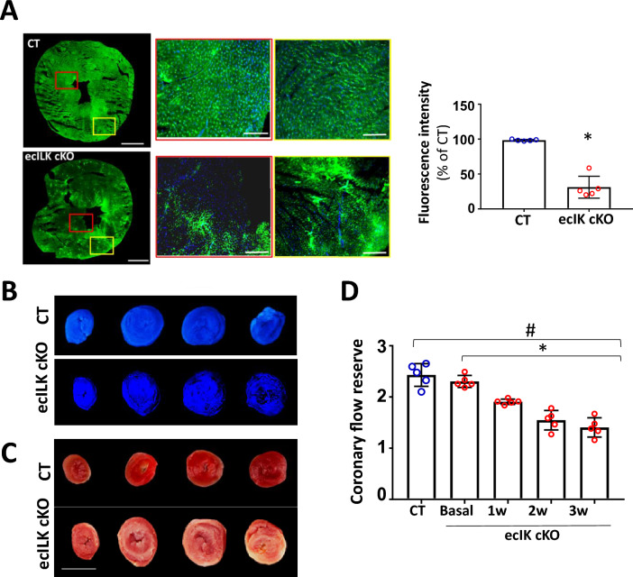
ILK deletion from coronary endothelium promoted microvascular dysfunction. A Representative confocal microscopy image of cardiac sections following in vivo dextran-fluorescein isothiocyanate (FITC) injection at 2 weeks after endothelial ILK deletion showing the absence of dextran-FITC (red box) and dextran-FITC accumulation (yellow box) in ecILK cKO microvessels compared to CT mice. Scale bar: 1 mm. Scale bar at magnifications: 100 mm. Right: fluorescence intensity quantitation expressed as percentage of CT (n = 6, * p < 0.005 vs CT). B and C Images show Thioflavin S and TTC staining in CT and ecILK cKO mice heart slices three weeks post deletion. B Under ultra-violet light, non-fluorescent regions on the Thioflavin S stain highlight areas of compromised coronary blood flow. (n = 4). C TTC sections indicate region of necrosis appearing white in the ecILK cKO group; CT group did no show infarction (n = 4). Scale bar = 5 mm. D Coronary flow reserve assessed by echocardiography at baseline and hyperemia in CT (3W) and ecILK cKO mice before (Basal) and at different time points after deletion showing decreased CFR at three weeks compared to CT and Basal (n = 5; *p < 0.001 vs Basal; #p < 0.001 vs CT). p values groups were assessed by ANOVA

