Fig. 7.
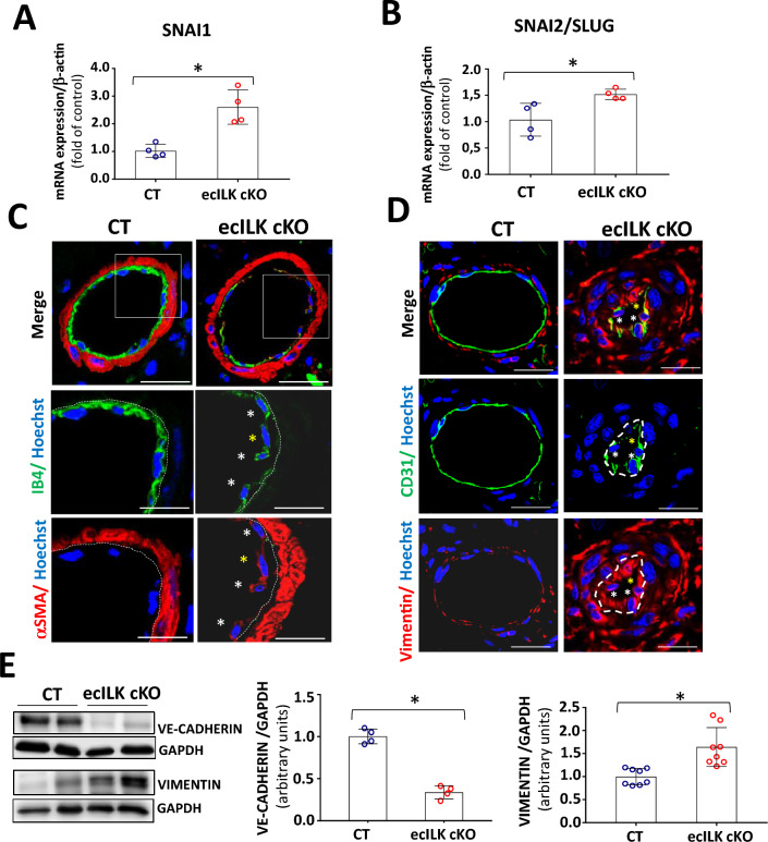
Endothelial ILK deletion induces endMT in coronary microvasculature. RT-qPCR of CT and ecILK cKO mice cardiac extracts showing mRNA expression of endMT transcription factors A Snai1 and B Snai2 (Slug) are significantly increased in ecILKcKO mice (*p < 0.05, n = 4). C Representative confocal photomicrographs showing arterioles in cardiac sections from CT and ecILK cKO mice immunostained for IB4 (green) and α-SMA (red). Nuclei were counterstained with Hoechst (blue). White asterisks mark endothelial cells expressing both endothelial and mesenchymal cell markers. Yellow asterisk mark an endothelial cell expressing mostly α-SMA. (Scale bar: 25 mm, n = 6). D Representative confocal photomicrographs showing small arterioles from CT and ecILK cKO mice immunostained for CD31 (green) and Vimentin (red). Nuclei were counterstained with Hoechst (blue). (Scale bar: 10 μm, n = 6). E Western blot analysis of VE-cadherin and Vimentin protein expression in CT and ecILK cKO cardiac protein extracts. GAPDH was used as a loading control. Left panel shows representative blots. Quantitative analysis shows a significant decrease of both proteins in ecILK cKO mice (*p < 0.05, n = 4–8 mice). p values were calculated using Student’s t test

