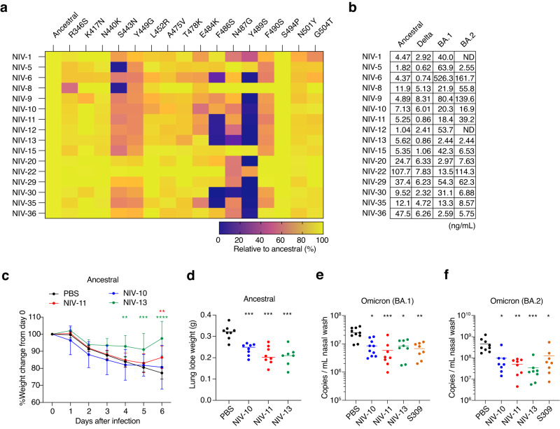
Fig. 2. Characterization of broadly neutralizing monoclonal antibodies.
a Binding profiles of each recombinant IgG1 clone to the corresponding RBD mutants were examined by mesoscale, and percentages relative to ancestral RBD are shown in color. b Neutralization of each recombinant antibody was examined with an authentic virus in vitro, and IC50 values are shown. ND, antibody without a detectable level of neutralization in the experimental condition. c, d In vivo treatment effect of indicated antibodies to Syrian hamsters infected with an ancestral virus. Syrian hamsters were intranasally inoculated with 104 TCID50 virus, and 5 mg/kg of each monoclonal antibody was administered intraperitoneally on the next day. Body weight change and weight of lung lobe on day 6 are shown. e, f In vivo treatment effect of indicated antibodies to Syrian hamsters infected with BA.1 (e) and BA.2 (f). Syrian hamsters were intranasally inoculated with 104 TCID50 virus, and 5 mg/kg of each monoclonal antibody was administered intraperitoneally on the next day. Subgenomic viral RNA copy numbers in the nasal wash on day 3 are shown. Data shown in a, b are representative of more than two independent experiments. Data shown in c–f are from two independent experiments. Data are presented as mean value ± SD (n = 8 biological replicate, c). Each symbol represents data from one hamster (n = 7–10 biological replicate, d–f), and bars represent the mean value (d–f). *p = <0.05 (e, f); **p = 0.004 (c, NIV-13), p = 0.0034 (c, NIV-11), p < 0.01 (e, f); ***p = 0.0006 (c), p < 0.001 (d–f); ****p < 0.0001 (two-way ANOVA with Dunnett’s test for body weight change, Kruskal–Wallis test for lung weight and qPCR).

