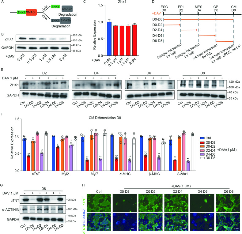
Fig. 2. Zhx1 specifically regulates the specification of CPs.
A The schematic diagram of SMASh-mediated ZHX1 degradation. B, C The protein (B) and mRNA (C) expression level of Zhx1 with the treatment of DAV in distinct concentrations (0, 0.5, 1, 1.5, and 2 μM). D A schematic diagram to represent the treatment timing and sample harvest during the SMASh-mediated ZHX1 knockdown. E The protein level of Zhx1 in the indicated days (D2, D4, D6, and D8) of cardiomyocyte differentiation after DAV treatment at different days (D0–D8, D0–D2, D2–D4, D4–D6, and D6–D8). F The expression of cardiomyocyte markers with the treatment of DAV (1 μM) at different days of cardiomyocyte differentiation. G The western blot results for the expression of cTNT and α-Actinin at day 8 of cardiomyocyte differentiation after DAV treatment (1 μM) at different days of cardiomyocyte differentiation. H The expression of cTNT protein with DAV treatment (1 μM) at different days of cardiomyocyte differentiation through immunofluorescent staining. Scale bar, 100 μm. Data are presented as the mean ± SEM (n = 3). The statistical significance is performed according to a two-way analysis of variance (ANOVA) followed by Bonferroni’s post hoc. *p < 0.05, **p < 0.01, and ***p < 0.001 versus Ctrl.

