Figure 1.
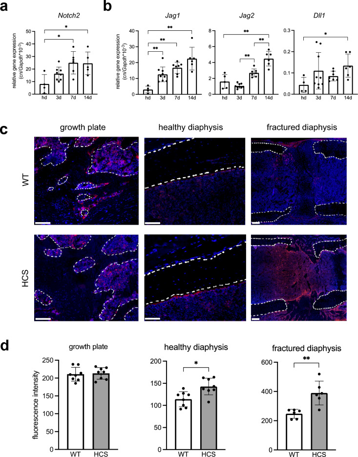
Notch2 is expressed in callus tissue. (a) Expression of Notch2 in the fracture callus of WT mice at the indicated time points. Hd = non-fractured, healthy diaphysis (b) Expression of the Notch receptor ligands Jagged1 (Jag1), Jagged2 (Jag2), and Delta-like-canonical-Notch-ligand-1 (Dll1) in the same samples. (c) Representative images of immunofluorescent stainings of Notch2 in the indicated femoral compartments. Upper row: non-fractured and fractured femur of WT mice. Lower row: non-fractured and fractured femur of HCS mice. Scale bar = 100 μm. Dotted white lines show trabecular bone (growth plate), non-fractured cortical bone (diaphysis), or fracture ends (diaphysis), respectively. (d) Quantification of respective immunofluorescent signal intensities of Notch2. For (a, b, d), non-parametric Mann–Whitney U test, *p < 0.05; **p < 0.005. Data are shown as mean with standard deviation. Data points indicate individual measurements from each mouse.

