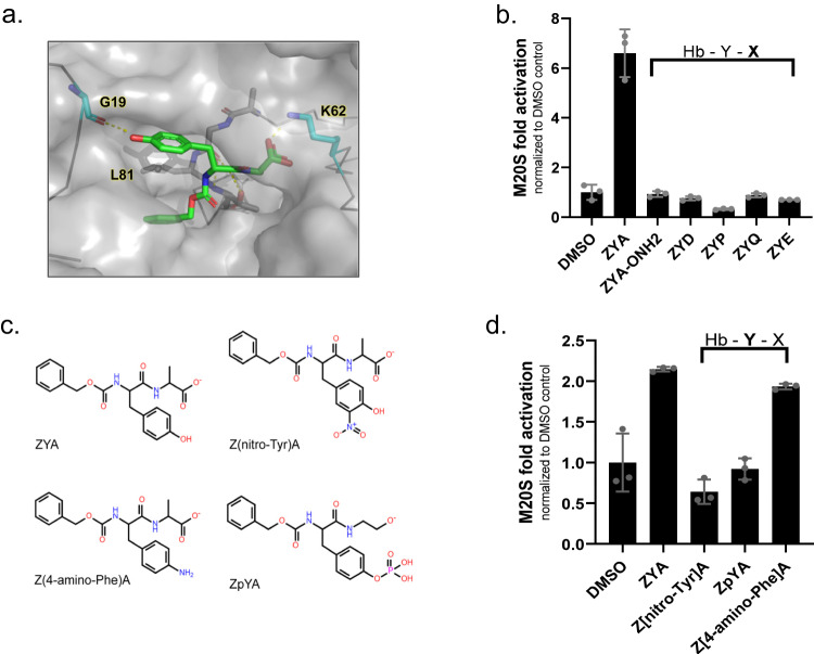
Fig. 5. Probing structure activity relationships of the ZYA gate-opening compound.
a ZYA (green sticks) docked in intersubunit pocket between α5&6 in human 20S. Image rendered in PyMOL. b Mammalian 20S proteasome (0.5 nM) activity (nLPnLD-amc hydrolysis, rfu/min) with the indicated ZYA derivatives (500 μM) with variations in the “X” position that were found to be deleterious to ZYA activity. Proteasome activity is normalized to DMSO. c Structures of derivatives tested in (d). d Mammalian 20S proteasome activity (nLPnLD-amc hydrolysis, rfu/min) with the indicated ZYA derivatives at a low binding concentration of 100 μM. Proteasome activity is normalized to DMSO. Data (means) are representative of three or more independent experiments each performed in triplicate. Error bars represent ± standard deviation.

