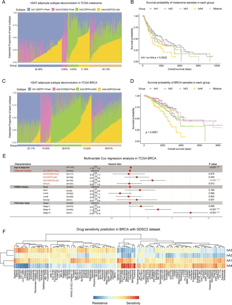
Fig. 6.
Deconvolution analyses in melanoma and BRCA cohorts. A Bar plot showing the estimated proportion of the three subcutaneous adipocyte subtypes in each TCGA melanoma sample. B Kaplan–Meier survival curve for TCGA melanoma cohort in five groups. P value was calculated with log‑rank test. Log‑rank p value < 0.05 was considered as statistically significant. C Bar plot showing the estimated proportion of the three subcutaneous adipocyte subtypes in each TCGA breast cancer sample. D Kaplan–Meier survival curve for TCGA breast cancer cohort in five groups. P value was calculated with log‑rank test. Log‑rank p value < 0.05 was considered as statistically significant. E Forest plots for multivariate regression of clinical factors and adipocyte subtypes in TCGA breast cancer datasets. F Pearson’s correlation of GDSC2 drug response (measured by IC50) with each of the four subcutaneous adipocyte subtype scores reveals drug resistance (blue) or sensitivity (red)

