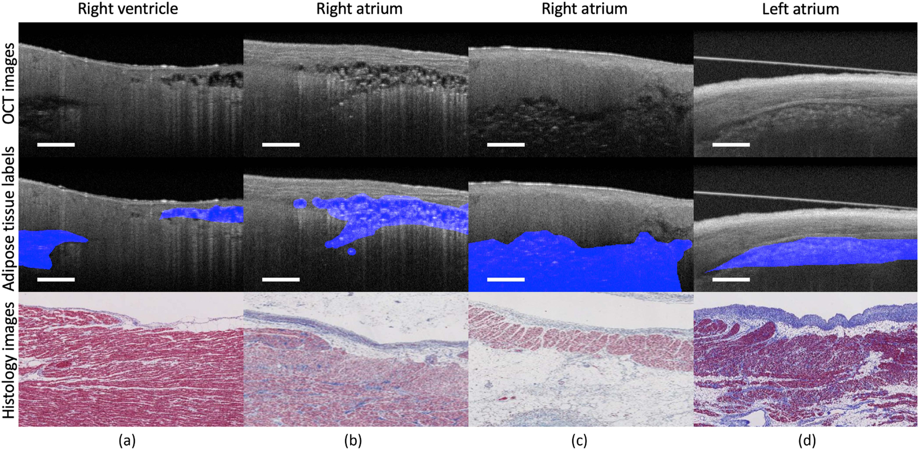
Fig. 1.

Representative OCT images from cardiac dataset. Sample (a) is obtained from right ventricle. Sample (b) and sample (c) are obtained from right atrium. Sample (d) is obtained from left atrium submerged in PBS solution. The features of adipose tissue present great variations among different locations and imaging conditions. The unclear boundary and irregular shape of adipose tissues add unique challenges for automated segmentation. Scale bar: 500 µm.
