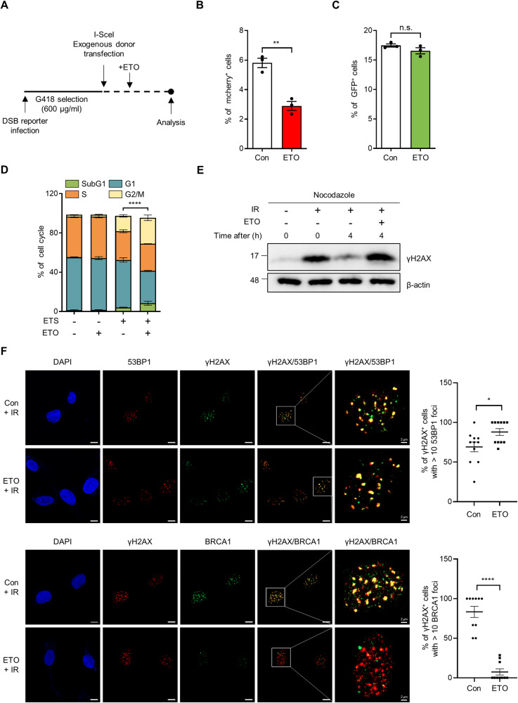
Fig. 2. FAO affects HR repair by promoting BRCA1 recruitment.
A Experimental design for Fig. 2B and C. 293 cells were infected with DSB reporter and selected with G418. Selected cells were transfected with exogenous donor and I-SceI. After transfection, cells were treated with or without ETO for 48 h and analyzed using FACS. Quantification of (B) mCherry and (C) GFP expression in BFP positive cells. Three million cells per sample were analyzed. Statistical analysis was based on two-tailed Student’s t-test. D Immortalized MEFs were treated with 0.5 μM ETS, 200 μM ETO, or both as indicated. The Cell cycle was analyzed by BrdU staining. Statistical analysis was performed using two-way ANOVA with Tukey’s multiple comparisons test. The indicated p-values (****) represent the comparative values between ETS-treated and ETS plus ETO-treated cells in G2/M phase. E γH2AX protein levels in immortalized MEFs. Cells, pre-treated with nocodazole (200 nM) for 20 h, were exposed to 3 Gy IR and then treated with or without 200 μM ETO for the indicated times. β-actin was used as loading control. F HeLa cells were exposed to 3 Gy IR and treated with or without ETO for 4 h. Representative images of immunofluorescence staining (53BP1, Red; γH2AX, Green; DAPI, Blue in upper panels and BRCA1, Green; γH2AX, Red; DAPI, Blue in lower panels). Scale bar represents 10 μm. Percentages of γH2AX positive cells with >10 Foci of BRCA1 or 53BP1 as indicated (right). Statistical analysis was performed using two-tailed Student’s t-test. All error bars ± SEM. *p < 0.05, **p < 0.01, and ****p < 0.0001.

