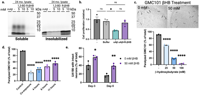
Fig. 4 |. β-hydroxybutyrate inhibits oligomeric toxicity and suppresses human amyloid-β-induced paralysis and neurotoxicity in C. elegans.
a, SDS-PAGE of HiLyte Fluor 488-labeled amyloid-β1–42 peptide monomer (mAβ) incubated with 24 month male wild-type soluble cytosolic brain proteins and treated with 5–10 mM of R-βHB and 5–10 mM of 1,3-BD. b, Quantification of N2a cell proliferation monitored by XTT Assay following treatment with 2 μM amyloid-β oligomers (oAβ) and 10 mM R-βHB. c-d, Quantification of amyloid-β proteotoxicity in temperature-sensitive (aggregation-permissive at +25°C) GMC101 strain, determined by scoring the percentage of animals paralyzed at (c) 25–28 hours following temperature shift and with 25–100 mM of βHB treatment (representative image shown), and (d) following temperature shift without treatment, then movement to 50 mM βHB treatment at varying timepoints. e, Quantification of amyloid-β neurotoxicity was determined by scoring number of intact glutaminergic neurons in UA198 animals (expressing amyloid-β in GFP-tagged glutaminergic neurons) with 50 mM of βHB treatment.
a, Representative image from triplicate repetitions.
b, Mean ± S.E.M, N=3, p-value calculated using one-way ANOVA with Tukey’s multiple comparison test.
c-d, Mean ± S.E.M, N=3 (~300 animals), p-value calculated using one-way ANOVA with Dunnett’s multiple comparison test.
e, Mean ± S.E.M, N=3 (~300 animals). p-value calculated using two-way ANOVA with Sidak’s multiple comparison test.

