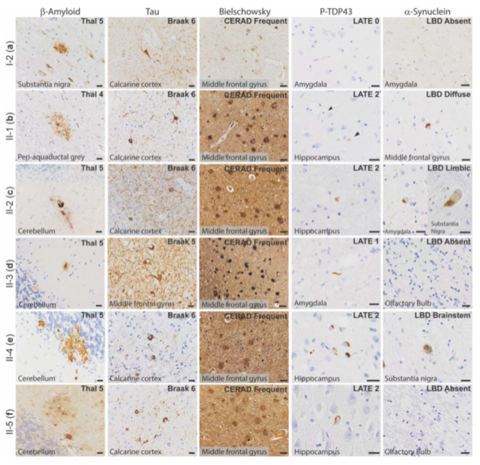
Figure 3. Neuropathology of all family members who consented to autopsy.
Representative photomicrographs demonstrating highest level neuropathologic change in each autopsy case for β-amyloid plaques (6e10 antibody), neurofibrillary tangles (tau antibodies as listed below), neuritic plaques (Bielschowsky silver stain), phosphorylated-TDP-43 inclusions (P-TDP43 antibody), and Lewy bodies (α-Synuclein antibody). (a) Patient I-2, with β-amyloid plaques in the substantia nigra, neurofibrillary tangles (Tau2 antibody) in the calcarine cortex (primary visual cortex), and frequent neuritic plaque density by CERAD criteria (note that silver staining was lighter than other cases). No p-TDP43 or α-synuclein was present, shown here as lack of staining in areas affected early in disease process. (b) Patient II-1, with β-amyloid plaques in the periaqueductal grey matter of the midbrain, neurofibrillary tangles (AT8 antibody) in the calcarine cortex, and frequent neuritic plaque density by CERAD criteria. P-TDP43 inclusions were present in the hippocampus, highlighted by arrows. Lewy bodies were present in brainstem, amygdala, limbic structures, and frontal cortex (shown here). (c) Patient II-2, with β-amyloid plaques in the cerebellum, neurofibrillary tangles in the calcarine cortex (AT8 antibody), and frequent neuritic plaque density by CERAD criteria. P-TDP43 inclusions were present in the hippocampus. Lewy bodies were present in the amygdala and substantia nigra, consistent with a limbic (transitional) pattern. (d) Patient II-3, with β-amyloid plaques in the cerebellum, neurofibrillary tangles in the middle frontal gyrus (AT8 antibody), and frequent neuritic plaque density by CERAD criteria. P-TDP43 inclusions were present in amygdala neurites. No Lewy bodies were observed, demonstrated here by negative staining of the olfactory bulb, one of the earliest anatomic sites of Lewy body formation. (e) Patient II-4, with β-amyloid plaques in the cerebellum, neurofibrillary tangles in the calcarine cortex (Tau2 antibody), and frequent neuritic plaque density by CERAD criteria. P-TDP43 inclusions were present in the hippocampus. Lewy bodies were present in the pigmented cells of the substantia nigra but not in any other site. (f) Patient II-5, with β-amyloid plaques in the cerebellum, neurofibrillary tangles in the calcarine cortex (Tau2 antibody), and frequent neuritic plaque density by CERAD criteria. P-TDP43 inclusions were present in the hippocampus. No Lewy bodies were observed, again demonstrated here by negative staining of the olfactory bulb. Scale bars = 20 μm for β-amyloid, p-tau, p-TDP43, and α-Synuclein; scale bars = 50 μm for Bielschowsky silver stain.

