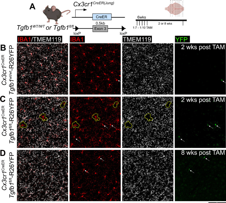
Figure 5. Sparse Tgfb1 induced-knockout in individual adult microglia supports an autocrine mechanism of microglial TGF-β ligand production and signaling regulation.
(A) Mouse model used to induce Tgfb1 KO in mosaic sparse individual microglia and experimental timeline depicting titrated dose of tamoxifen. (B-D) Representative images showing IBA1, TMEM119, and YFP expression and co-localization in control tissue at 2 weeks post tamoxifen (B) and sparse iKO tissue at 2 weeks (C) showing loss of TMEM119 expression in sparse individual microglia and (D) reversal of TMEM119 expression in the sparse Tgfb1 iKO brain at 8 weeks post tamoxifen. The yellow dotted outline in (C) highlights singular microglia showing loss of homeostatic TMEM119 expression. White arrows highlight YFP+ cells showing no loss of homeostatic TMEM119 expression. Note that at this low dosage of TAM, the recombination of individual floxed alleles (R26-YFP reporter or the floxed Tgfb1 gene) occurs independently of each other, therefore YFP+ cells do not always indicate a sparse Tgfb1 KO microglia, consistent with our recent study25. Representative results from n=3–5 mice/group from different cohorts of TAM treatment. Scale bar = 100µm.

