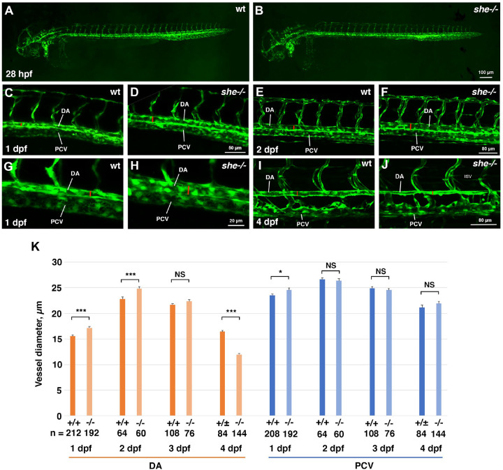
Figure 2. she mutants show enlarged diameter of the dorsal aorta.
(A,B) Overall vascular patterning of she−/− mutants is normal when compared to their sibling wild-type embryos at 28 hpf. Embryos are in kdrl:GFP background.
(C-H) A wider DA is observed in she mutant embryos compared to their wild-type (she+/+) siblings at 1 and 2 dpf (28 and 48 hpf respectively). Red line indicates DA diameter. (G,H) shows higher magnification image of trunk vasculature at 1 dpf.
(I,J) DA is narrower in she mutants at 4 dpf compared to their siblings.
(K) Diameter of the DA and PCV at 1–4 dpf in she mutants and their wild-type siblings. * p<0.05; ***p<0.001, NS – not significant, Student t-test. Error bars show SEM.
At all stages, she mutant and sibling embryos were obtained by in-crossing sheci26+/−; kdrl:GFP carriers. Embryos at 1 and 2 dpf were genotyped after imaging. Embryos at 4 dpf were separated based on the phenotype, and wild-type siblings include she+/+ and she+/− embryos at this stage.

