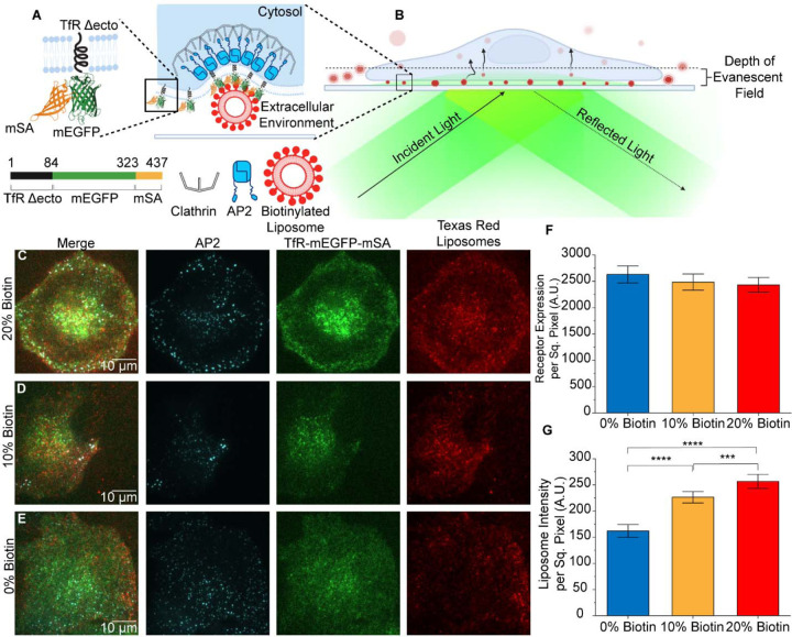
Figure 1. Targeting promotes penetration of liposomes beneath adherent cells.
A. The chimeric receptor, TfR-mEGFP-mSA, which was designed to recruit biotinylated liposomes to endocytic sites. B. Schematic of TIRF microscopy to examine internalization of liposomes by endocytosis from the adherent surfaces of cells. C-E. TIRF microscopy images of AP2 (JF646), chimeric receptor (EGFP), and liposomes (Texas Red DHPE) at the adherent surfaces of SUM159 cells. All channels have been contrasted equally across all three conditions. F. Average fluorescence intensity of the chimeric receptor on the plasma membrane. No significant differences were seen between groups of cells exposed to each population of liposomes. (t-test 0% vs 10%; p = 0.514; n = 107, 182) t-test 0% vs 20%; p = 0.353; n = 107; 166; t-test 10% vs 20%; p = 0.800; n = 182, 166). G. Total fluorescence intensity per area of liposomes present beneath cells. (t-test 0% vs 10%; p < 1×10⁻⁴; n = 107, 182) t-test 0% vs 20%; p < 1×10⁻⁴; n = 107; 166; t-test 10% vs 20%; p = 3.55×10⁻⁴; n = 182, 166). For F and G, three independent trials were acquired for each condition with a minimum of 15 cells imaged per trial. Error bars represent the standard error of the mean, where N is the number of cellular crops analyzed across all trials. Significance between conditions was identified using a two-tailed student’s t-test and a one-factor ANOVA with α = 0.05.

