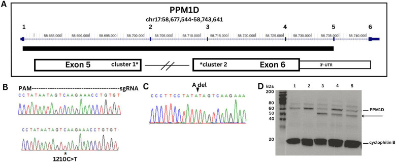
Figure 1. DNA sequence analysis and Western blot of PPM1D truncating variants.
A. Map of PPM1D showing the 6 exons with the catalytic domain depicted as a solid bar. The two clusters of JdVS mutations are in the 3'-end of exon 5 and the 5'-end of exon 6. B. DNA sequence strip of wild type allele on top and the patient sample showing the c.1210C>T; p.Q404X nonsense mutation on bottom. The region covered by the guide RNA used for CRISPR-Cas 9 engineering is shown. C. Two isogenic lines with an "A" deletion were generated with CRISPR. D. PPM1D Western blot showing wild-type protein and the truncated protein. Cyclophilin is a loading control. Lanes 1 and 2 are control samples, lane 3 is the patient sample, and lanes 4 and 5 are two CRISPR lines.

