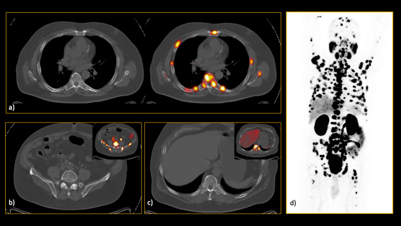
Fig. 2.
In this interesting case, a 66-year-old man referred for initial staging, there was extensive involvement of both axial and appendicular skeleton. However, none of the lesions had significant characteristics on the low-dose CT images, resulting in calling this patient free of skeletal involvement in CT evaluation by all readers. As you can see in the hybrid 68Ga-PSMA PET/CT images, there were innumerable bone metastases which all were invisible on CT alone (a–c). d The maximum intensity projection (MIP) clearly visualises the extent of the disease

