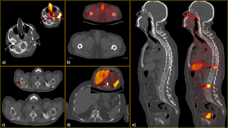
Fig. 3.
False-negative low-dose CT findings (red arrows). In each section (a–e), the CT-alone images, along with the hybrid 68Ga-PSMA PET/CT images, can be found. Regarding section a), it should be noted that he was one of our interesting cases, as he showed only a single bone metastasis in the skull base. Thus, although the sclerotic lesion can be easily seen in this single slice, it was not prominent enough while scrolling, since the remainder of the skeleton was free of metastases. Furthermore, in section c), subtle sclerosis could be found retrospectively; however, it was not considered a significant lesion by all readers when reviewing CT images. Notably, this false finding did not misclassify the patient’s disease burden eventually

