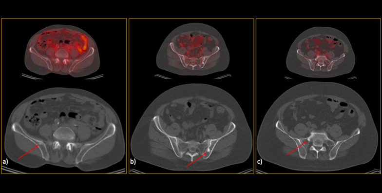
Fig. 4.
False-positive low-dose CT findings (red arrows). The majority of the false-positive findings were located in the pelvic region, most likely due to the readers’ knowledge about the high pretest probability in this region. So, as can be seen (a–c), there were various lesions called metastatic on CT images but were found benign considering the reference standard. Noteworthy, although b and c might seem not that much challenging and in favour of bone islands while looking at only one slice, in patients with multiple metastases, they were falsely interpreted as malignant since there were also other lesions with more or less similar Hounsfield unit in the same patient

